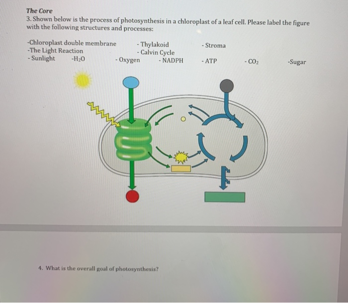
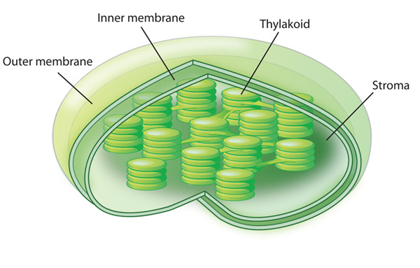
43 label the chloroplast in the figure below
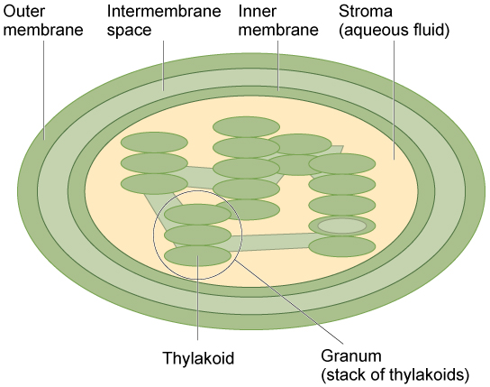
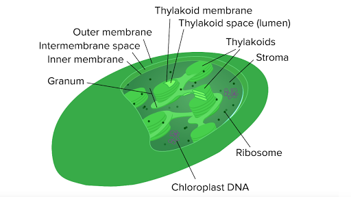
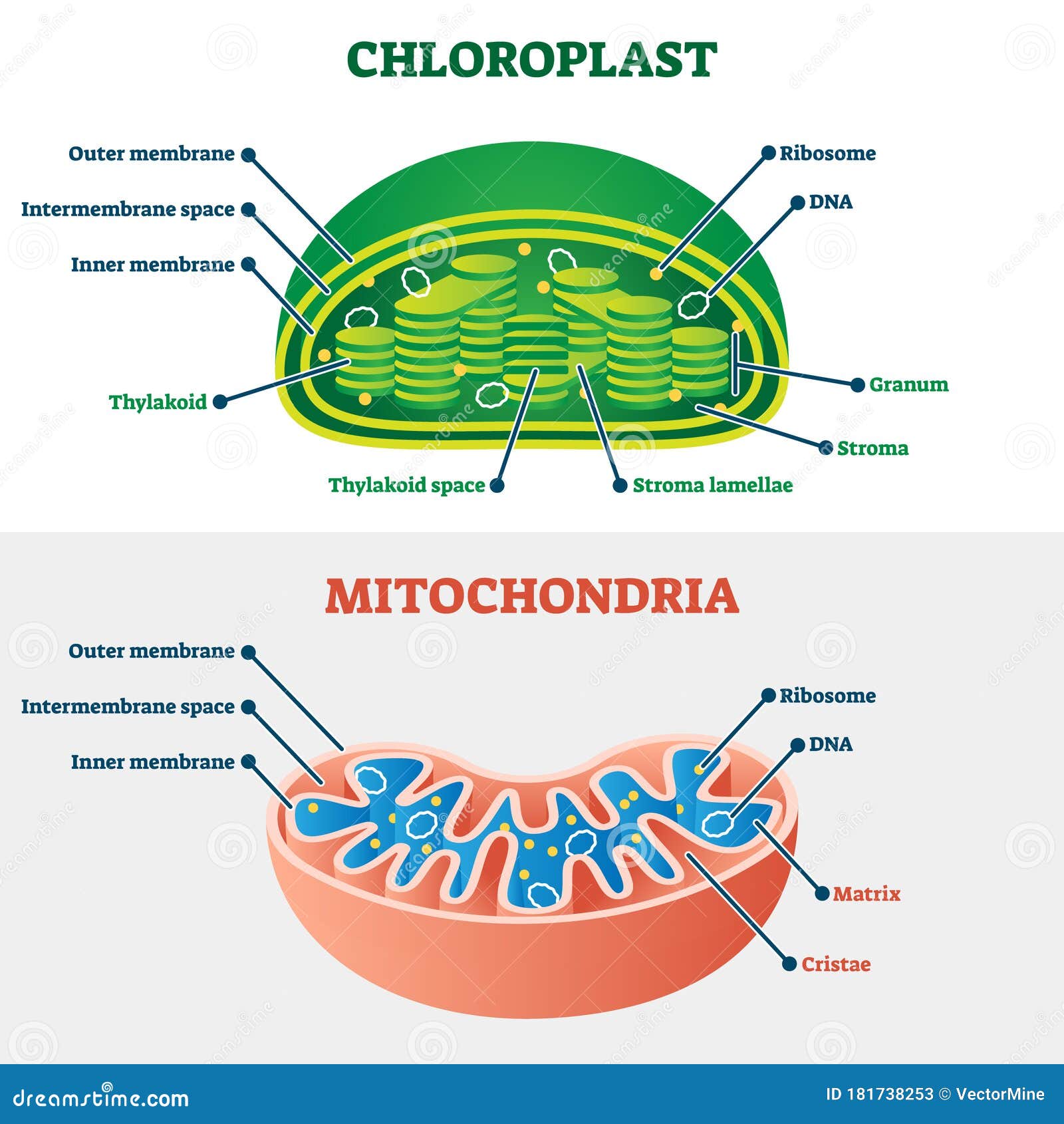
Chloroplast Labels Diagram | Quizlet Protects and holds the form of the organelle. Stroma Transparent fluid. Thylakoid A disc-shaped structure inside a chloroplast where the light-dependent reactions of photosynthesis take place Granum A stack of thylakoids in a chloroplast Lamellae Tubes that connect the granum Inner membrane The location of the electron transport system Organism - Definition and Examples - Biology Online Dictionary 15.06.2022 · An organism refers to a living thing that has an organized structure, can react to stimuli, reproduce, grow, adapt, and maintain homeostasis.An organism would, therefore, be any animal, plant, fungus, protist, bacterium, or archaeon on earth.These organisms may be classified in various ways. One of the ways is by basing upon the number of cells that make it up.
Lab 2 Cells Worksheet.docx - Lab #2 Worksheet - Course Hero Onions do not have chloroplasts because they grow underground with no light. 6. Describe the shape and the location of chloroplasts. They are round, oval or disk shaped. They are located in all green tissues, mostly in the leaf's parenchyma cells. 2. Biasing from the model predictions will be prevented because of imputation. 3. TargetBuy

Label the chloroplast in the figure below
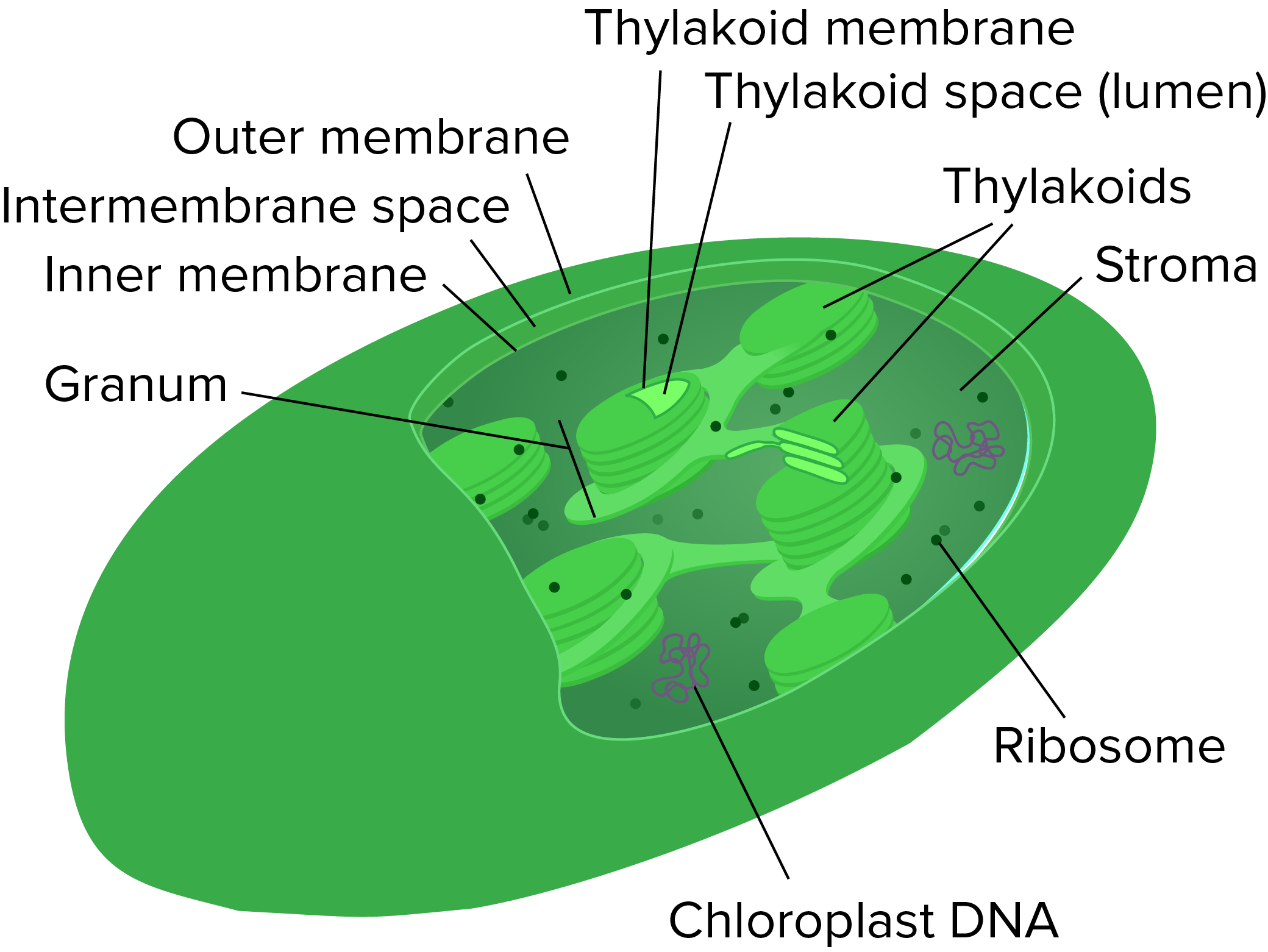
Solved Label the chloroplast below: Include the following - Chegg Transcribed image text: Label the chloroplast below: Include the following structures: Thylakoid, granum, stroma, thylakoid space, inner membrane, intermembrane space, outer membrane: Figure 1 WOW I The general principle is that the green parts of plants can use light to produce organic compounds and oxygen from carbon dioxide and water. Write the formula below: What photosynthesis actually ... ScienceDirect - A single-cell Arabidopsis root atlas reveals ... 08.02.2022 · Download : Download video (13MB) Video S1. Animation showing 3D UMAP of the atlas with cell type annotations, related to Figure 1.. The Arabidopsis root is a tractable model organ with established markers for most cell types as well as expression profiles for morphologically defined developmental stages (Birnbaum et al., 2003; Brady et al., 2007a; Li et … Chloroplast: Meaning, Structure, Analogy - Embibe The diagram of Chloroplast is given below. Fig: A Labeled Diagram of Chloroplast Chloroplast Structure Chloroplasts are roughly \ (1 - 2\, {\rm {μm}}\) thick and \ (5 - 7\, {\rm {μm}}\) in diameter and are seen in all higher plants. In different plants, chloroplasts have different shapes like some plants have filamentous or ovoid or saucer-shaped.
Label the chloroplast in the figure below. Chloroplast: Structure and Function - Biology Wise Structure. Chloroplasts are located in the parenchyma cells of plants as well as in autotrophic algae. They are oval-shaped organelles having a diameter of 2 - 10 µm and a thickness of 1 - 2 µm. Although their dimensions are almost similar in all plants, the algal chloroplasts show a variation in their size as well as shape. ICSE Solutions for Class 10 Biology – Photosynthesis 04.12.2019 · Sketch and Label the Diagram Question 1: Draw a neat and well-labeled diagram of the Chloroplast. Answer: Question 2: Draw a neat and well-labeled diagram of the apparatus you would set up to show that oxygen is given out during photosynthesis. Answer: See figure. Explain the Terms. Question: 1. ATP 2. Calvin Cycle 3. Free Energy 4. NADPH 5 ... A Golgi-localized Hexose Transporter Is Involved in Heterotrimeric … Figure 7 F is a three-dimensional reconstruction from a Z-stack acquisition of control and BFA-treated cells. Bodipy-BFA has been previously shown to label the Golgi complex in living cells (Deng et al., 1995). We used Bodipy-BFA to stain Golgi in Arabidopsis seedlings expressing SGB1- GFP driven by the cauliflower mosaic virus (CaMV) 35S promoter. DOC Read, Answer, Color, Label: Chloroplasts - New Smyrna Beach High School Chloroplasts are double-membrane organelles with a smooth outer membrane and an inner membrane folded into disk-shaped sacs called thylakoids. Color and label the outer membrane (1) green. Color and label the inner membrane (5) yellow. Thylakoids, containing chlorophyll and other accessory pigments
Presenting Data – Graphs and Tables – Principles of Biology The structure of the chloroplast. Light and Pigments. The Light-Dependent Reactions. ... Label the X and Y axis, including units. Graph your data. Add a descriptive caption to your graph. Note that data tables are titled above the figure and graphs are captioned below the figure. Solved Chloroplast structure Label the chloroplast in the | Chegg.com Expert Answer The given image is of a chlorop … View the full answer Transcribed image text: Chloroplast structure Label the chloroplast in the figure below. Thylakoid Outer membrane Inner membrane Grana Thylakoid space Stroma Outer membrane E Thylakoid Previous question Next question Biology Test #1 Flashcards | Quizlet Match each label with the correct structure in the figure, structure of mitochondria. ... Match each label with the correct structure in the figure, structure of chloroplasts.-stroma- fluid matrix inside the chloroplasts-granum- the stack of thylakoids ... Complete the sentences below by making use of the periodic table.-Bromine has (35 ... PDF Chloroplasts and Mitochondria - Mrs. Slovacek's Science In the figure below: 1. Color and label the outer membrane light green. 2. Color and label the grana (STACK) dark green. 3. Color and label the lamellae brown. 4. Color and label the stroma light blue. 5. Label a single thylakoid and a single granum. FIGURE 1-CHLOROPLAST Outer Membrane Grana Lamellae Stroma Thylakoid Granum 2
The structure of the chloroplast - Principles of Biology Chloroplasts are surrounded by a double membrane similar to the double membrane found within a mitochondrion. Within the chloroplast is a third membrane that forms stacked, disc-shaped structures called thylakoids. Embedded in the thylakoid membrane are molecules of chlorophyll, a pigment (a molecule that absorbs light) through which the entire ... Chapter 26, Problem 53P | bartleby Ch. 26 - Figure 26-16 shows cross sections through three... Ch. 26 - Figure 26-17 shows a rectangular solid conductor... Ch. 26 - Figure 26-18 shows plots of the current i through... Ch. 26 - Figure 26-19 shows four situations in which... Ch. 26 - In Fig. 26-20, a wire that carries a current... Ch. 26 - Figure 26-21 gives the electric potential ... Organism - Definition and Examples - Biology Online Dictionary Jun 15, 2022 · The label determines where the protein would next go, i.e. for transport outside or within the cell. The mitochondrion (plural: mitochondria) is a semi-autonomous organelle that is responsible for generating ATP (by citric acid cycle and oxidative phosphorylation pathways). It is a semi-autonomous organelle because it has its own genetic material. Chloroplast- Diagram, Structure and Function Of Chloroplast - BYJUS Chloroplasts are found in all higher plants. It is oval or biconvex, found within the mesophyll of the plant cell. The size of the chloroplast usually varies between 4-6 µm in diameter and 1-3 µm in thickness. They are double-membrane organelle with the presence of outer, inner and intermembrane space.
DOC Read, Answer, Color, Label: Mitochondria - New Smyrna Beach High School Color and label. the outer membrane (8) purple and the cristae (9) red on Figure 3. The cristae greatly increase the surface area of the membrane so that the carbohydrates (simple sugars) can combine with oxygen to produce ATP (adenosine triphosphate). The electron transport chain takes place across the membranes of the cristae (9) (crista ...
Chapter 26, Problem 53P | bartleby Ch. 26 - Figure 26-16 shows cross sections through three... Ch. 26 - Figure 26-17 shows a rectangular solid conductor... Ch. 26 - Figure 26-18 shows plots of the current i through... Ch. 26 - Figure 26-19 shows four situations in which... Ch. 26 - In Fig. 26-20, a wire that carries a current... Ch. 26 - Figure 26-21 gives the electric potential ...
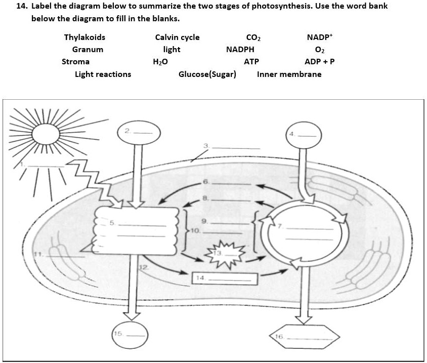
ICSE Solutions for Class 10 Biology – Photosynthesis Dec 04, 2019 · Sketch and Label the Diagram Question 1: Draw a neat and well-labeled diagram of the Chloroplast. Answer: Question 2: Draw a neat and well-labeled diagram of the apparatus you would set up to show that oxygen is given out during photosynthesis. Answer: See figure. Explain the Terms. Question: 1. ATP 2. Calvin Cycle 3. Free Energy 4. NADPH 5 ...
Extra Questions for Class 9 Science Chapter 5 The Fundamental ... (a) Chloroplast – Containing chlorophyll (b) Chromoplast – Containing carotenoids and xanthophyll (coloured plastids) (c) Leucoplast – White or colourless plastids 2: What is plasma membrane made up of? Answer: Plasma membrane is made up of proteins and lipids. 3: What did Robert Hooke observed first in cork cell?
PDF Biology 29 Cell Structure and Function Spring, 2009 Springer - Amherst Remember to properly label the axes of the graphs and to include proper units. Each figure should have a legend: beginning with (1) a figure number, followed by (2) a descriptive title (an incomplete sentence) specific to your data, and then (3) the rest of the legend in complete sentences. The legend is found at the bottom of the figure.
(PDF) Raven Biology of Plants | Michael Branks - Academia.edu Enter the email address you signed up with and we'll email you a reset link.
Group 1 Killing Chloroplasts Worksheet.docx - Names:... Here are five tables, one for each of the five herbicides being tested, showing data on NADPH production (nmol/g) in chloroplasts in the absence of herbicide (Before) and 10 minutes after addition of herbicide (After). Analyze the data and then draw a graph to represent the findings. The first herbicide (H 1) is done for you and ‐ there is a graph started. As appropriate, you will want to ...
Chapter 3 homework (study guide) Flashcards | Quizlet Fill out the chart using the labels. Some labels may be used more than once. Label the image below to assess your understanding of prokaryotic cell structure. Indicate if the following statements about prokaryotes are true or false. A. They contain a plasma membrane. True B. They contain internal membrane-bound organelles such as a nucleus. False
Plasmid - Wikipedia In order for plasmids to replicate independently within a cell, they must possess a stretch of DNA that can act as an origin of replication.The self-replicating unit, in this case, the plasmid, is called a replicon.A typical bacterial replicon may consist of a number of elements, such as the gene for plasmid-specific replication initiation protein (Rep), repeating units called iterons, DnaA ...
Presenting Data – Graphs and Tables – Principles of Biology Notice that the graph has a descriptive caption that allows the figure to stand alone without additional information given from the procedure: you know that this graph shows the average of the measurements taken from four tomato plants. Figure 4: Average growth (in cm) of tomato plants when treated with different brands of fertilizer.
Academia.edu - Cambridge International AS and A Level Biology ... • Metabolism: all the chemical processes occurring within an organism • Enzymes increase the rate of reactions that occur in living organisms.
chloroplast | Definition, Function, Structure, Location, & Diagram A chloroplast is a type of plastid (a saclike organelle with a double membrane) that serves as the site of photosynthesis, the process by which energy from the Sun is converted into chemical energy for growth. Chloroplasts contain the pigment chlorophyll to absorb light energy.
The structure labeled as X is the - Brainly.com Answer: Label X is Nucleus ; Eukayotic cell ; Label W is Chloroplast Explanation: The given image is a eukayotic cell as it has well defined nucleus and membrane bound cell organelles such as nucleus, mitochondria, chloroplast.. The label X is nucleus, It contains genetic material and it controls all the activities of the cell.. The label W is chloroplast.
NCERT Class 10 Science Lab Manual Stomata - CBSE Tuts In the figure given below of stomata the correct label for A and B are (a) A - nucleus, B - guard cells (b) A - guard cells, B - nucleus (c) A - chloroplast, B - nucleus (d) A- chloroplast, B - guard cell. . Question 14: The temporary mount of stomata was seen under the microscope which appeared pink in colour. The stain used was ...
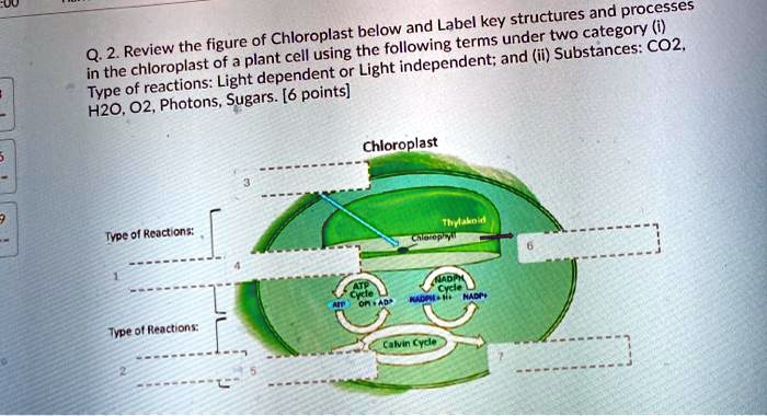
PDF 198 Chloroplasts SF - CXC® CAPE® Biology Resources For Caribbean Students Which organelle is the chloroplast? (1) 2. The diagram below shows the structure of a chloroplast, as seen using the electron microscope. B C A (a) Name the parts labelled A, B and C.(3) (b) Give the letter of the structure where (i) the light-dependent reactions of photosynthesis take place (1) (ii) the light-independent reactions take place (1)
Plasmid - Wikipedia History. The term plasmid was introduced in 1952 by the American molecular biologist Joshua Lederberg to refer to "any extrachromosomal hereditary determinant." The term's early usage included any bacterial genetic material that exists extrachromosomally for at least part of its replication cycle, but because that description includes bacterial viruses, the notion of plasmid was refined over ...
(PDF) Raven Biology of Plants | Michael Branks - Academia.edu Enter the email address you signed up with and we'll email you a reset link.
Label the diagram below i cell wall ii plasma membrane iii nucleus iv ... Place the usedtoothpick (s) in the container provided. Identify the cell membrane, cytoplasm, and nucleus in individual cells. Draw one epithelial cell in the space below and label the above structures. Draw the cell at least 4cm in diameter and indicate the magnification of the drawing (as you did with the onion cell).
Miko Barrios BIO15 Lab 6.pdf - Photosynthesis Lab Learning... View Miko Barrios BIO15 Lab 6.pdf from BIO 15 at Solano Community College. Photosynthesis Lab Learning Objectives • • • • • • Describe the anatomy of a chloroplast. Know where each stage of
Mastering Biology Chapter 4 Flashcards | Quizlet The figure below shows a Paramecium, a single-celled freshwater protist. ... Drag the labels onto the diagram to identify the structures of an animal cell. a. cytoskeleton b. ribosomes c. nucleus d. smooth endoplasmic reticulum (ER) ... Chloroplast - makes food by converting light energy into chemical energy
BIO Chapter 4 Quiz Flashcards | Quizlet Label the chloroplast in the figure below.-Match each description with the correct organelle: mitochondria, chloroplasts, or both. Mitochondria: Extract energy from food Site of cellular respiration ... Label the figure below to assess your knowledge of osmosis in plant and animal cells. Place the labels describing the type of solution into the ...
Structure of Chloroplast (With Diagram) | Botany - Biology Discussion 1. Chloroplasts (Figs. 295-296), responsible for the photosynthesis of the plants, are the characteristic features of the cells of green plants. ADVERTISEMENTS: 2. Around the chloroplast is present a double membrane envelope. 3. Each membrane of chloroplasst is 35 to 50 Å thick. 4. Many dark-coloured grana are present.
Chloroplasts- Definition, Structure, Functions and Diagram - Microbe Notes Chloroplasts are a type of membrane-bound plastids that contain a network of membranes embedded into a liquid matrix and harbor the photosynthetic pigment called chlorophyll. It is this pigment that imparts a green color to plant parts and serves to capture light energy. Chloroplasts can be found in the cells of the mesophyll in plant leaves.
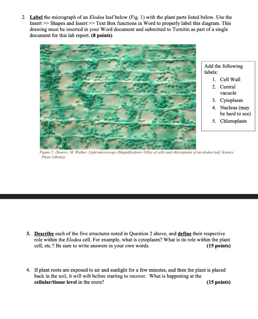
Chloroplasts contain coloured pigments. Figure given below shows some ... Chloroplasts contain coloured pigments. Figure given below shows some of the cells from a leaf that contains chloroplasts. Make a large drawing to show the three complete cells containing chloroplasts in above figure. Label one chloroplast on your drawing. Difficulty - medium Solving time: 3 mins Need help understanding this concept?
Chloroplasts - Structure And Functions - A Level Biology In algae a single huge chloroplast is seen that appears as a network, a spiral band or a stellate plate. The size of the chloroplast also varies from species to species and it is constant for a given cell type. In higher plants, the average size of chloroplast is 4-6 µ in diameter and 1-3 µ in thickness. Parts of Chloroplasts
Learn Insta - Life Processes Class 10 Important Questions and Answers ... 03.08.2020 · (a) Stomata. They are pores regualted by two chloroplast containing guard cells that occur in the epidermis of leaves and young stems for exchange of gases and transpiration. (b) Diagram. (c) Functions of Stomata, Exchange of gases, Transpiration. Question 5. (a) Name the process by which autotrophs prepare their own food.
Chloroplast: Meaning, Structure, Analogy - Embibe The diagram of Chloroplast is given below. Fig: A Labeled Diagram of Chloroplast Chloroplast Structure Chloroplasts are roughly \ (1 - 2\, {\rm {μm}}\) thick and \ (5 - 7\, {\rm {μm}}\) in diameter and are seen in all higher plants. In different plants, chloroplasts have different shapes like some plants have filamentous or ovoid or saucer-shaped.
ScienceDirect - A single-cell Arabidopsis root atlas reveals ... 08.02.2022 · Download : Download video (13MB) Video S1. Animation showing 3D UMAP of the atlas with cell type annotations, related to Figure 1.. The Arabidopsis root is a tractable model organ with established markers for most cell types as well as expression profiles for morphologically defined developmental stages (Birnbaum et al., 2003; Brady et al., 2007a; Li et …
Solved Label the chloroplast below: Include the following - Chegg Transcribed image text: Label the chloroplast below: Include the following structures: Thylakoid, granum, stroma, thylakoid space, inner membrane, intermembrane space, outer membrane: Figure 1 WOW I The general principle is that the green parts of plants can use light to produce organic compounds and oxygen from carbon dioxide and water. Write the formula below: What photosynthesis actually ...
















Komentar
Posting Komentar