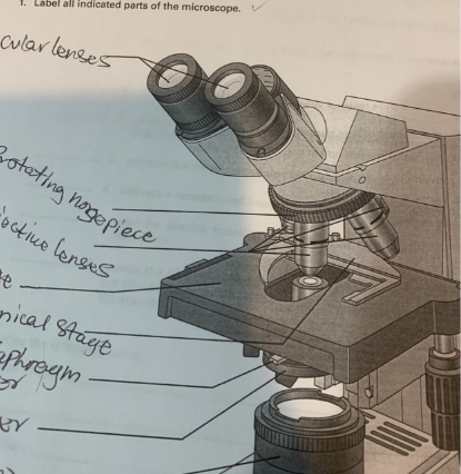
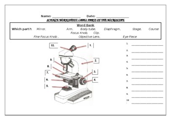
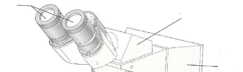
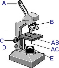
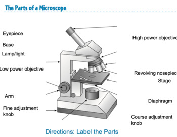
44 label the indicated parts of the microscope
Lab 1- The Microscope- Lab Report 3 - Using the picture, label the ... Lab 1- The Microscope- Lab Report 3 - Using the picture, label the indicated parts of the microscope - Studocu lab lab the lab report using the picture, label the indicated parts of the microscope with the terms listed in the blue box. each term is used only once. switch Skip to document Ask an Expert Sign inRegister Sign inRegister Home Parts of the Microscope with Labeling (also Free Printouts) A microscope is one of the invaluable tools in the laboratory setting. It is used to observe things that cannot be seen by the naked eye. Table of Contents 1. Eyepiece 2. Body tube/Head 3. Turret/Nose piece 4. Objective lenses 5. Knobs (fine and coarse) 6. Stage and stage clips 7. Aperture 9. Condenser 10. Condenser focus knob 11. Iris diaphragm
Label the microscope — Science Learning Hub In this interactive, you can label the different parts of a microscope. Use this with the Microscope parts activity to help students identify and label the main parts of a microscope and then describe their functions. Drag and drop the text labels onto the microscope diagram.

Label the indicated parts of the microscope
Exercise 3: The Microscope Flashcards - Easy Notecards label all indicated parts of the microscope. 6. explain the proper technique for transporting the microscope. when transporting the microscope, hold it in an upright position with one hand on its arm and the other supporting its base. avoid swinging the instrument during its transport and jarring the instrument when setting it down. Lab 1: The Microscopic World - Biology LibreTexts Open combination drawer and take out the microscope. 3. Label all the parts of the microscope with the provided post-its using the image below or the laboratory manual. Note. The image below does not match your microscope perfectly, you will be responsible for knowing the parts of your microscope on the lab practical. Label the parts of the microscope below in the corresponding numbers ... Label the parts of the microscope below in the corresponding numbers under the. Label the parts of the microscope below in the. School Suffern Senior High School; Course Title SCIENCE REG; Uploaded By mia23reyna23. Pages 4 This preview shows page 3 - 4 out of 4 pages.
Label the indicated parts of the microscope. Label parts on microscope Flashcards | Quizlet Eyepiece Body Tube on microscope High power objective Low power objective Stage Opening Diaphragm Lever Light on microscope Coarse adjustment Fine adjustment Nosepiece Arm on a microscope Stage Clip Stage on microscope Base Students also viewed Realidades 1B 45 terms Images senoramix Teacher Protists 8 terms MsCleary83 Teacher Solved PART D: Assessments 1. Label the microscope parts in | Chegg.com Answer : Label Number Parts Description 1 Body Tube (Eye piece tube) Passes light from the head to the eyepiece. They connects eyepiece to the objective lenses. 2 Rotating Head Contains mirrors … View the full answer Transcribed image text: PART D: Assessments 1. Label the microscope parts in figure 4.8. Microscope Parts, Types & Diagram | What is a Microscope? The major parts of a microscope include the head, arm, base, and stage. Other parts include the eyepiece and objective lenses. What are microscopes used for? One use for a microscope is to... 3.1: Introduction to the Microscope - Biology LibreTexts The optical system of a compound microscope consists of two lens systems: one found in the objective (s) lens (es) (Fig. 2, part 3); the other in the ocular (eyepiece) (Fig. 2 part 1). The objective lens system is found attached to a rotating nosepiece (Fig. 2, part 2).
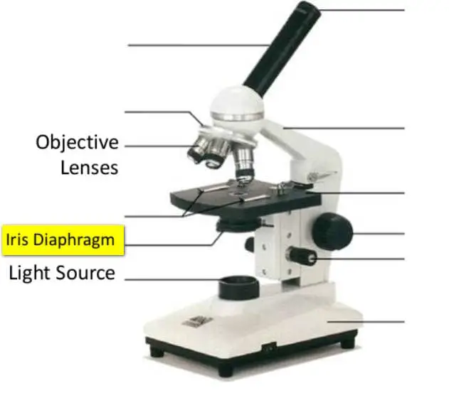
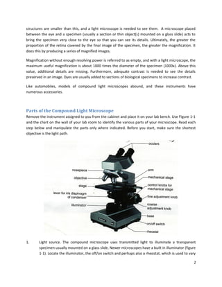
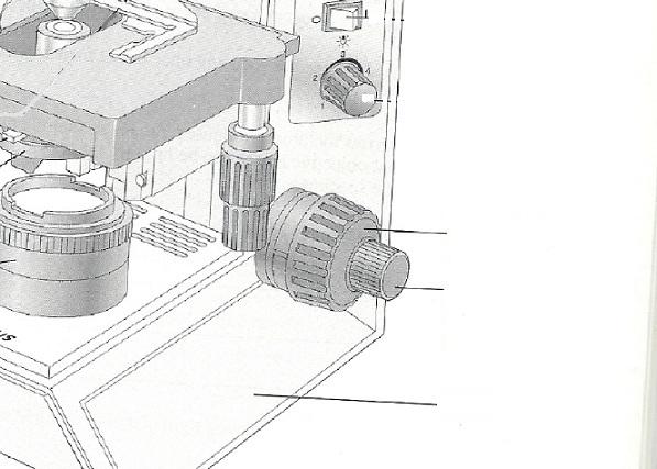
Parts of a Microscope with Their Functions • Microbe Online Parts of Compound Microscope Illuminator (Light Source) Diaphragm (Iris) Condenser Aperture Stage Objective lens Body Tube Ocular Lens (eye-piece) Coarse and Fine Adjustment Knob Arm Base Microscope Worksheet The Light Microscope Light microscopes are used to examine cells at relatively low magnifications. Parts of the Microscope (Labeled Diagrams) It is also known as bright-field microscope because it enables the light to pass directly through the source of light through the two lenses. Let us discuss the different parts of a compound microscope. a. Mechanical Parts of a Compound Microscope Foot or Base. The base is a U-shaped structure that bears all of the compound microscope weight. PDF Label parts of the Microscope Label parts of the Microscope: . Created Date: 20150715115425Z 3.1: Examining epithelial tissue under the microscope 1. Plug in the microscope & turn on light source. 2. Pick up microscope by carrying arm, position it so it is accessible to your seat, with open side of the stage facing you. 3. Rotate the objectives so that the lowest power objective (smallest in size) clicks into place. 4. Look at the slide with your naked eye and find the location of the ...
Labeling the Parts of the Microscope | Microscope World Resources Labeling the Parts of the Microscope This activity has been designed for use in homes and schools. Each microscope layout (both blank and the version with answers) are available as PDF downloads. You can view a more in-depth review of each part of the microscope here. Download the Label the Parts of the Microscope PDF printable version here. Label all indicated parts of the microscope. | Quizlet Label all indicated parts of the microscope. Solution. Verified. Answered 1 year ago. Answered 1 year ago. Step 1. ... objective lens; Step 2. 2 of 3. The middle part of the microscope from top to the bottom): mechanical stage (ensures the microscope slide) stage; condenser (for focusing the light on the specimen) iris diaphragm lever (for ... Molecular Expressions Microscopy Primer: Anatomy of the Microscope ... Microscope manufacturers offer a wide range of objective designs to meet the performance needs of specialized imaging methods, to compensate for cover glass thickness variations, and to increase the effective working distance of the objective. Learn to identify microscope objectives and their specialized properties by deciphering the engravings on the barrel. Microscope: Parts Of A Microscope With Functions And Labeled Diagram. The microscope has three basic components: the head, the base, and the arm. Head:Occasionally, the head is considered the body. It holds the optical components of the upper part of the microscope. Base:The microscope's base provides great support. It is also equipped with miniature illuminators.
MICRODRIVE AND TELEMETRY SYSTEM FOR MULTI-UNIT EXTRACELLULAR ...
Parts of a microscope with functions and labeled diagram - Microbe Notes There are three structural parts of the microscope i.e. head, base, and arm. Head - This is also known as the body. It carries the optical parts in the upper part of the microscope. Base - It acts as microscopes support. It also carries microscopic illuminators.

List: Parts of a Microscope and their Function | Pathwooded
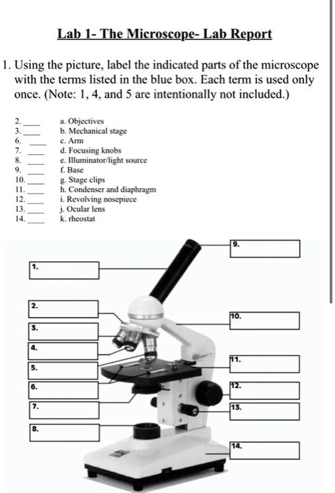
Solved Lab 1- The Microscope-Lab Report 1. Using the - Chegg Using the picture, label the indicated parts of the microscope with the terms listed in the blue box. Each term is used only once. (Note: 1, 4, and 5 are intentionally not included.) 2. 3. 6. 7. 8. 9. 10. 11. 12. 13. 14. a. Objectives b. Mechanical stage c. Arm d. Focusing knobs e. Illuminator/light source f. Base g. Stage clips h.

Microscope With Labels clip art | Microscope parts, Science ...
Label the parts of the microscope below in the corresponding numbers ... Label the parts of the microscope below in the corresponding numbers under the. Label the parts of the microscope below in the. School Suffern Senior High School; Course Title SCIENCE REG; Uploaded By mia23reyna23. Pages 4 This preview shows page 3 - 4 out of 4 pages.

Thrombus Structural Composition in Cardiovascular Disease ...
Lab 1: The Microscopic World - Biology LibreTexts Open combination drawer and take out the microscope. 3. Label all the parts of the microscope with the provided post-its using the image below or the laboratory manual. Note. The image below does not match your microscope perfectly, you will be responsible for knowing the parts of your microscope on the lab practical.
Parts of the Microscope with Labeling (also Free Printouts ...
Exercise 3: The Microscope Flashcards - Easy Notecards label all indicated parts of the microscope. 6. explain the proper technique for transporting the microscope. when transporting the microscope, hold it in an upright position with one hand on its arm and the other supporting its base. avoid swinging the instrument during its transport and jarring the instrument when setting it down.

Label Microscope Diagram - EnchantedLearning.com

Portable handheld two-photon microscope imaging of a skin ...
Microscope Parts Quiz
SOLVED: Lab LTheMicroscgpe-Lab Report Using the picture ...

Parts of Stereo Microscope (Dissecting microscope) – labeled ...

Parts of a microscope with functions and labeled diagram

PART 1. SCIENTIFIC METHOD I. STEPS - ppt download
Solved) - Care and Structure of the Compound Microscope 1 ...
1.5: Microscopy - Biology LibreTexts

Compound Microscope Parts, Functions, and Labeled Diagram ...
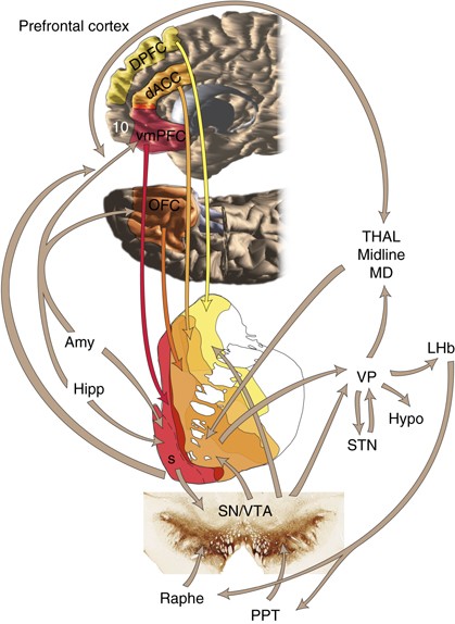
The Reward Circuit: Linking Primate Anatomy and Human Imaging ...
Parts Of The Microscope Label Teaching Resources | TPT
Example test image from the 2D Electron Microscopy Cell ...
Activity 1 Review of Microscopy

Lab Book - RS 3.pdf - NAME LAB I'IME'DATE The Microscope Care ...
Parts of the Microscope with Labeling (also Free Printouts ...
Multifocal imaging for precise, label-free tracking of fast ...
Exercise 3: The Microscope Flashcards - Easy Notecards
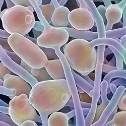
Fungi | What is microbiology? | Microbiology Society

SOLUTION: Label the Parts of a Microscope - Studypool
Microscope Quiz
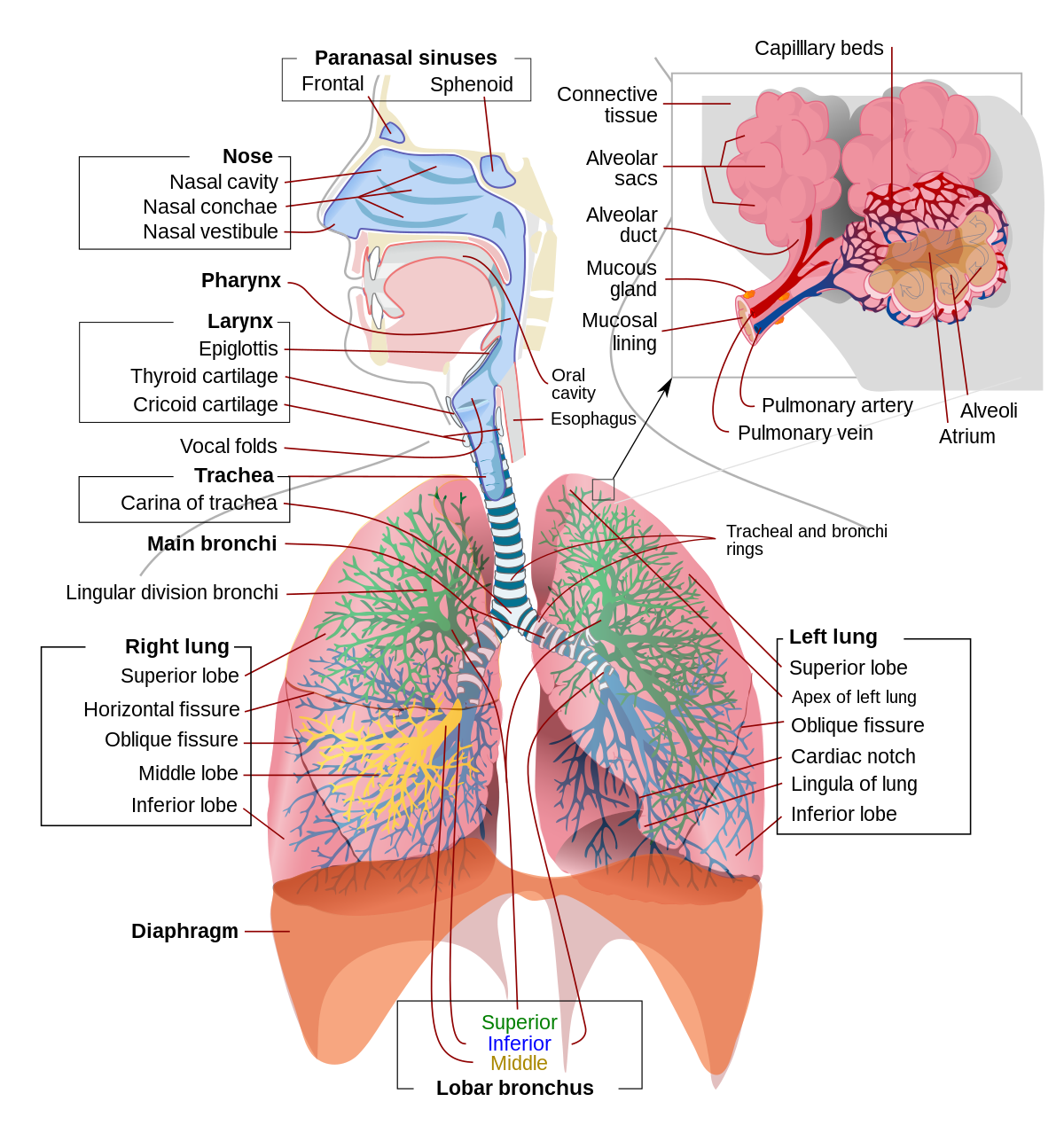
Respiratory system - Wikipedia

Step-by-step preparation of mouse eye sections for routine ...

morphology Flashcards | Quizlet
Anatomy of organs of the digestive system and their functions ...
Brainstem: Definition, anatomy, parts, function | Kenhub

Types, Parts and Functions of a Microscope

Parts of a Microscope Diagram | Quizlet
Microscopy 1

Parts of a Microscope - SmartSchool Systems

Lab Chapter #2 - The Microscope Diagram | Quizlet

Compound Microscope Parts – Labeled Diagram and their ...
Exercise 3: The Microscope Flashcards - Easy Notecards
An Introduction to the Light Microscope, Light Microscopy ...

Mention the Function of Each Microscope Part - BYJU'S Biology
Microscope Bundle!! - Parts of a Microscope Unit Activities
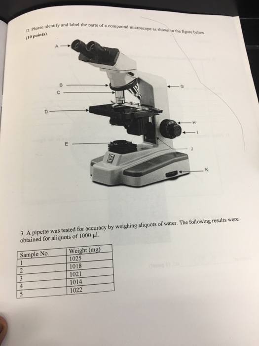
Solved Identify and label the parts of a compound microscope ...

SOLUTION: Label the Parts of a Microscope - Studypool

PPT - Label the parts on your microscope picture. PowerPoint ...


Komentar
Posting Komentar